THE SIGNAL IN ONE SENTENCE
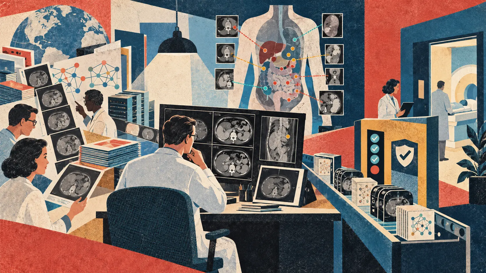
Alibaba DAMO Academy and a large hospital and university research group have released RADAR, a vision-language model built to examine contrast-enhanced abdominal CT scans across 18 anatomical structures and 146 imaging findings. The peer-reviewed paper appeared in Science on September 17. The public repository provides training, preprocessing and inference code under Apache 2.0, while the downloadable model checkpoints and auxiliary data use the more restrictive CC BY-NC-SA 4.0 license. That split matters. Researchers can inspect and modify the software, and they can download weights for non-commercial work under the model terms, but this is not a permission slip to install RADAR in a hospital and start making treatment decisions. The paper reports training on more than 400,000 examinations and 15 million anatomy-wise image-text pairs. Its abstract says the model generalized across multiple centers and that assistance improved the diagnostic sensitivity of 26 radiologists by about 10 percent. Those are serious research results. They are not regulatory clearance, a prospective clinical trial, universal superiority to doctors or proof that every one of 146 findings works equally well. The plain signal is that the lab door is open wider than usual. The clinic door still has a regulator, a local validation team, a security review and a radiologist standing in front of it, which is exactly where they belong.
01
WHAT ACTUALLY CHANGED
The RADAR team published An expert-level generalist AI for abdominal CT diagnosis in Science on September 17. PubMed records the paper, its authors and affiliations across Alibaba DAMO Academy, Zhejiang University, Chinese hospitals and international research institutions. The work is centered in Hangzhou and a broad clinical network rather than being a repackaged United States product launch.
RADAR is designed as a generalist model for contrast-enhanced abdominal computed tomography. Instead of training a separate detector for one disease, the team paired image regions with anatomy-level text from clinical reports. The paper abstract reports coverage of 18 anatomical structures and 146 imaging findings. That means 146 categories of radiological findings, not 146 cancers and not 146 complete patient diagnoses.
The training scale is large for this domain. The authors report more than 400,000 CT examinations and 15 million anatomy-wise image-text pairs. The model learned from existing clinical reports without manual annotation of every finding. That reduces labeling cost, but it also means the system can inherit the omissions, wording habits and institutional biases present in those reports.
The paper reports internal and external evaluation across multiple centers and varied scenarios. Its abstract says RADAR achieved high diagnostic performance and robust generalization. The public inference guide gives one reproducible slice of that claim: on the external MERLIN test set, the released checkpoint produced an average area under the receiver operating characteristic curve of 0.883 across 21 listed findings, with individual values ranging from 0.6834 for fracture to 0.9903 for abdominal aortic aneurysm.
A reader study involved 26 radiologists. The paper abstract says RADAR assistance increased their diagnostic sensitivity by about 10 percent. Sensitivity asks how many true positive findings were caught. It does not by itself tell us how many false alarms appeared, whether reading quality improved for every radiologist, or whether patients experienced better outcomes.
The release is more substantial than a screenshot and a press quote. The GitHub repository includes code for training, inference and preprocessing, documentation, result files and download scripts. The model page lists the pretrained RADAR checkpoint, Chinese and English text encoders, a vision checkpoint and two RADAR+ checkpoints. The inference guide says a demonstration can run on one A100 or H20 graphics processor.
The word open needs a footnote. The repository code declares Apache 2.0, a permissive software license. The Hugging Face model card labels the checkpoints CC BY-NC-SA 4.0, which requires attribution, restricts commercial use and applies share-alike terms. The auxiliary anatomical-mask data uses the same non-commercial license. Code, weights and data are available, but they do not all carry the same freedoms.
No reviewed source identifies a regulatory clearance, approved clinical indication, commercial hospital deployment, prospective patient trial or autonomous-use authorization for RADAR. The public materials describe a research model and evaluation workflow. Calling it a released diagnostic product would skip the most important part of the story.
02
WHY THIS MATTERS
Abdominal CT is an unusually crowded visual task. One scan can contain urgent disease, chronic findings, incidental abnormalities, treatment effects and normal variation across many organs. A model that looks broadly rather than waiting for a single requested target could help radiologists avoid tunnel vision, especially when the original clinical question points attention in the wrong direction.
The reader study points toward the most plausible near-term role: a second pass. A radiologist reads the scan, the model raises candidate findings, and the human decides what belongs in the report. That is less cinematic than an autonomous robot doctor and much more useful. It can also fail in quieter ways, such as automation bias, alert fatigue or a tendency to chase every highlighted speck.
Breadth changes the validation problem. A tool for one narrow finding can be evaluated against one clinical use case. A 146-finding system needs evidence across common and rare conditions, positive and negative cases, scanners, contrast protocols, hospitals, demographic groups and levels of disease severity. A good average can hide a weak tail, and the weak tail is where an uncommon emergency may be sitting.
External validation is better than testing only where a model was trained. It asks whether performance survives a change in institution and data. It is still not the same as prospective deployment. Once the system enters a live workflow, patient mix shifts, reports are incomplete, images arrive with artifacts, staff use the interface in unexpected ways and the model may change how radiologists search the scan.
The public release makes scrutiny possible. Independent teams can inspect preprocessing assumptions, reproduce the MERLIN evaluation, test different populations and look for failure clusters. That is a meaningful scientific advantage over a closed demonstration. Reproducibility will still depend on access to suitable CT data, specialist labels, capable hardware and the exact licensed components.
License clarity matters for hospitals and startups. Apache 2.0 code can support broad reuse, but non-commercial model weights cannot simply be folded into a paid clinical service. A team must read every component license, including upstream text encoders, anatomical tools and data sets, then determine whether a planned use is permitted. Open on a download page is not the same as commercially deployable.
The model learned supervision from clinical reports rather than perfectly annotated ground truth. That is clever and scalable. It can also reproduce what earlier radiologists tended to mention, underreport or phrase ambiguously. A finding absent from a report is not always absent from the scan. The training method therefore deserves auditing alongside the network itself.
China has a large clinical imaging ecosystem and a strong incentive to stretch scarce specialist time. A broad model developed with Chinese hospitals could be locally important and globally instructive. Translation is not automatic. Protocols, disease prevalence, reporting culture, equipment, regulation and liability vary by country, so each health system still needs evidence on its own patients and workflow.
03
WHERE IT COULD HELP
- Use RADAR as a research benchmark for broad abdominal CT finding detection, with per-finding results instead of one flattering average
- Run retrospective local validation on a held-out hospital cohort before any live workflow test, stratified by site, scanner, protocol, age, sex and clinically relevant patient groups
- Study human plus model performance by measuring missed findings, false positives, reading time, report changes, disagreement patterns and whether radiologists become over-reliant on suggestions
- Build a second-reader workflow that keeps the original scan, model output, final report and human rationale linked for audit without turning every low-confidence score into an interruptive alert
- Test distribution shift across emergency, outpatient, oncology, postoperative and screening populations rather than assuming one result represents every abdominal CT service
- Create a monitored research deployment with versioned weights, access controls, protected health information safeguards, drift checks, rollback procedures and incident reporting before considering clinical scale
- Audit the anatomy-level report supervision for missing labels, ambiguous phrases and institutional habits, then compare model errors with the weaknesses of the source reports
- Review code, weight, data and dependency licenses separately before adapting or commercializing the stack, and document which component authorizes each planned use
KEEP A HAND ON THE WHEEL
RADAR has peer-reviewed evidence and a serious public research release, but the reviewed materials do not establish regulatory approval, prospective clinical safety, patient-outcome benefit, commercial-use permission for the weights or dependable performance in every country and hospital. The headline number of 146 refers to imaging findings, not 146 cancers or 146 final diagnoses. Area under the curve measures ranking discrimination across thresholds. It is not accuracy, a calibrated probability, a clinical decision, a false-alarm count or an outcome measure. The public external MERLIN example covers 21 listed findings and reports an average AUC of 0.883, with substantial variation by finding. The reader-study sensitivity gain does not by itself disclose specificity, all subgroup effects, every reader result or the effect of repeated daily use. Contrast-enhanced abdominal CT is also only one imaging context. Watch for the complete paper methods and supplements, prospective multicenter trials, independent replications, regulatory filings, per-finding calibration, subgroup and protocol results, false-positive burden, security and privacy reviews, interface studies, post-deployment monitoring and a clean explanation of which licenses apply to code, checkpoints and data.
04
TERMS WORTH KEEPING
SOURCES AND VERIFICATION STATUS
This article was written from the materials below. Product claims and dates were checked against those sources on September 19, 2026.
PUBLICATION RECEIPT: Revision 1. Published September 19, 2026.
THE PUBLICATION ENGINE
WANT A SIGNAL OF YOUR OWN?
We build source-grounded publications, private briefings, and editorial systems for organizations with something useful to say.
WORK WITH US